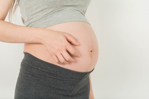
Pregnancy Cholestasis
There is severe itching in pregnancy cholestasis that occurs in the second half of pregnancy. It is related to the high levels of bile acids in the mother’s blood and increased risks for the unborn baby. It is the most common pregnancy-related liver disease. It was first described by Ahlfeld in 1883.
Its frequency varies according to geographical regions. While it varies between 1 / 100000-3 / 1000 in the United States, it is seen in the frequency of 7/1000 in England and 1-2 / 100 in Scandinavia. While the frequency is 15% in Chile, its frequency is low in America, Sweden and France.
When the frequency increases;
* A history of cholestasis in previous pregnancy
* Twin pregnancies (20-22%)
* Pregnancies caused by assisted reproductive techniques (2.7%)
* Pregnancy over 35 years old
* Presence of hepatitis C
* The presence of a gallstone history in the woman herself and her family.
The causes of pregnancy cholestasis are explained by the interaction of multiple factors. Here, genetic, hormonal, environmental and nutritional factors can contribute to cholestasis.
Nutrition
Selenium deficiency in nutrition is thought to increase the frequency of cholestasis. Selenium; It acts as a cofactor with functional effects for most of the liver enzymes, and deficiency may produce problems with bile formation and secretion.
Genetic Factors
A number of gene mutations that affect carrier proteins in the bile ducts can affect the transport of bile acids and cause their accumulation.
In 50% of women with cholestasis in geographical regions with high frequency, cholestasis is present in family history. This supports the autosomal dominant transition in genetic transmission.
Hormonal Factors
There is a lot of natural evidence to suggest that pregnancy cholestasis may be related to estrogen and progesterone hormone levels. Here, in the second half of pregnancy, the increase in the frequency of multiple pregnancies and the repetition of bile acid height in pregnant women who had gestational cholestasis afterwards are used. All of these hormones are metabolized in the liver, and the products that appear after metabolism are thought to affect bile duct movements. There are also publications that think that the effect of progesterone is more than estrogen.
The rapid disappearance of symptoms after pregnancy also supports the presence of hormonal causes.
Findings and Laboratory Assessments
The main finding is pruritus and usually occurs before laboratory findings. Itching can be widespread all day and as well, more often at night it can be seen intensely on the palms and soles of the palms. It usually occurs in the second trimester. Pruritus may also occur in the first trimester as hormone activity is high in multiple pregnancies. Jaundice occurs in only 10% of itchy pregnant women.
Hepatic function tests (transaminases) are found high in tests performed after itching.
The most sensitive marker in the diagnosis of pregnancy cholestasis is the height detected in total bile acids in serum. Apart from that, transaminases, alkaline phosphatase, gamma glutamyl transferase and bilirubins are also evaluated in the laboratory. Transaminases, called ALT and AST, increased by 2-10 folds or more. In 40% of patients, ALT may increase from 10 folds more. However, in 50% of patients, gammaglutamile transferase value increases. Also, bilirubin values can be found high in 10-20% of patients.
In differential diagnosis, viral hepatitis, HELLP Syndrome and acute fatty liver of pregnancy should be excluded. Because in this disease, the monitoring of the mother and the fetus, the course and risks of the diseases and the management of the diseases will be completely different.
Maternal Factors
In pregnancy cholestasis, a few days after the pregnancy is terminated, liver enzymes decrease and complaints are reduced. If liver enzyme elevation persists after pregnancy, this is no longer related to gestational cholestasis and other liver diseases that may lie below should be investigated.
It should be kept in mind that cholestasis may recur in the use of birth control pills after birth.
In women who had gestational cholestasis, the probability of having cholestasis in the next pregnancy was given as 45-70%.
Fetal Factors
In pregnancy cholestasis, the effects of the fetus are more important and may have bad results compared to the mother.
Problems such as increase in meconium amniotic fluid, oxygenation problems in the mother’s womb, fetal distress, premature labour and sudden loss of baby are more common in cholestasis compared to normal pregnancies.
Poor progress in pregnancy is observed with a probability of 90% after 37 weeks. Therefore, termination of pregnancy in pregnancies with cholestasis 37 weeks and earlier has reduced the poor fetal outcomes in recent years.
Bad fetal outcomes in pregnancy are directly proportional to the severity of gestational cholestasis. Fetal risks are less common if the total serum bile acids level is below 40nmol / L.
The frequency of encounter with meconium-containing amniotic fluid in the normal course of pregnancy was reported as 15%, this rate was found to be around 16-58% in cholestasis.
Although the mechanism of premature birth is not fully known, oxytocin receptor activity is known to increase in pregnancy cholestasis. Itching and high detected bile acids should be accepted as precursors of premature labour.
While the frequency of sudden baby loss in the womb is 10-15%, it is 3.5% today. Here, ursodeoxycholic acid therapy, frequent fetal follow-up (with NST and ultrasonography), more frequent biochemical analysis, and termination of pregnancies at 37-38 weeks significantly reduced fetal loss rates.
Treatment
The purpose of treatment in pregnancy cholestasis is to decrease the itching of the mother and to improve the laboratory findings and to decrease the risks of the fetus.
Although many agents have been tried in the treatment, the most promising treatment is seen as ursodeoxycholic acid. Cholestyramine can reduce maternal effects, but it cannot provide favourable effects in terms of fetal effects. At the same time, cholestyramine may increase nausea, vomiting and bleeding tendency in the mother and baby due to vitamin K absorption deficiency.
Ursodeoxycholic acid is a natural bile acid that makes up 5% of bile acids in humans. There is no obvious side effect of the mother or fetus. Its effect is realized through three mechanisms. The first mechanism is to reduce bile acid release. In this way, bile acid and bilirubin levels detected in maternal blood decrease. Two other mechanisms are the protection of bile duct cells and liver cells from the toxic effects of bile acids. All of these therapeutic properties seem to be superior to other treatment options.
Effects related to the fetus are controversial. Although there are reports showing that premature birth, meconium amniotic fluid and fetal distress frequency decrease with ursodeoxycholic acid, there is no clear evidence that fetal effects have improved yet. There was no improvement in fetal effects in drug options other than ursodeoxycholic acid.
Follow-up of pregnancy in pregnancy cholestasis and purpose of treatment; mother’s complaints are reduced and the comfort of the mother, premature birth of baby, reduction of fetal distress and meconium amniotic fluid rates.
When pregnancy cholestasis is diagnosed, routine initiation of ursodeoxycholic acid is the accepted treatment option.
Although a standard way is not recommended in the follow-up of pregnancy, it should be perceived that pregnancy detected with cholestasis is now a high risk pregnancy. For this reason, fetus is evaluated more frequently by ultrasonography and non-stress test (NST) is applied during pregnancy follow-up. The risks of premature birth and the risk of loss in the womb should be evaluated separately for each case, and appropriate delivery timing should be decided together with laboratory data and fetus data. We know that NST and fetal biophysical profile monitoring is unlikely to predict the risk of sudden death of babies in the womb. In this sense, follow-up and tests cannot predict the possibility of sudden death of the fetus. Sudden loss of the fetus is often due to acute oxygen deficiency. In these babies, there are no signs of chronic oxygen deficiency. Fetuses frequently gain weight and grow as in normal pregnancies, and umbilical artery doppler values are often found normal.
Since fetal death is frequently seen at 37 weeks and later, it is necessary to plan the birth around 37-38 weeks in women with gestational cholestasis. Since it may be necessary to consider premature birth at the border, the timing of birth should be calculated individually by calculating the risks.


 Turkish
 Deutsch
 Bu İçeriği Beğendim