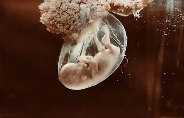
Location Abnormalities of Placenta
Abnormalities related to placement of the placenta in the intrauterine tissue, which is the main center of circulation between mother and baby, are important causes of postpartum hemorrhage.
These are under the main headings;
* placenta previa, where the placenta is located in the lower segment, that is, in the cervix,
*placenta accreta, placenta increata, and placenta percreata, which express the adhesion disorders of the placenta to the uterine muscle wall,
* ablation placenta, which means premature separation of the placenta,
* much more rarely observed placenta bilobata, placenta membranacea and accessory lobe.
Placenta Previa
Placenta 24-28 of pregnancy. It is defined as the partial or complete placement of the uterus in the lower segment, which is the exit of the uterus after the week.
The classification is all about how much the placenta closes the cervix.
1) Placenta previa totalis: The cervix is completely closed by the placenta. It is the heaviest form.
2) Placenta previa partial: The cervix is partially closed with the placenta.
3) Placenta previa marginalis: The border of the placenta is at the edge of the cervix entrance, but does not cover the entrance.
4) Down-placed placenta: The edge of the placenta does not reach the cervix but is very close.
Plasenta previa yaklaşık olarak 300 doğumda bir görülmektedir.
There are a number of risk factors that increase the previa frequency:
1) Having a previa in previous pregnancy increases the risk 12 times.
2) The woman had previous caesarean section; increases the risk in proportion to the number of caesarean. While the probability of previa is 0.65% in a woman who has previously been cesarean, the risk reaches around 1.5% after the second cesarean, 2.2% with the third cesarean and 10% with the fourth cesarean.
3) As the number of births of women increases, the probability of previa increases.
4) Advanced maternal age is interpreted as a risk factor for previa. However, the effect of muscle tissue sensitivity or number of births should not be ignored.
5) Increased placental surface in multiple pregnancies increases the possibility of previa.
6) Finally, due to the increase in carbon monoxide in cigarette smoking, the previa frequency increases by increasing the surface area of the placenta and increasing oxygen uptake.
The most common clinical finding in diagnosis; painless vaginal bleeding. Bleeding is generally not common before the end of the first three months. Repeater is monitored as non-excessive bleeding. Rarely, severe bleeding may occur from the beginning. In order for bleeding to risk fetus life, there must be severe bleeding that will shock the mother. Manual examination of the abdominal area is not very guiding. Only when the lower surface of the uterus is palpated by hand can it be doubted that the part that has not entered the pelvis has not entered. However, the diagnosis cannot be made on the basis of clinical bases.
Ultrasonography is the gold standard in diagnosis. Timing of ultrasonography in diagnosis is very important. While ultrasonography performed before the 24th week of gestation was followed by 28% of the placentae, after 24th week, the rate drops to 18% in term, and that is 3% at the end of pregnancy. It should be remembered that the population frequency of Previa is 1/300.
Vaginal ultrasonography is much more sensitive for the diagnosis of Previa and, more importantly, the rating of localization, and should be evaluated with vaginal ultrasonography in suspected cases. It will provide a clearer diagnosis especially in placenta from the back wall. 20-23. If there is a placenta that closes at least 25 mm cervix in weeks, vaginal delivery at term is not possible. 32-34 at lesser values. In weeks, vaginal ultrasonography clarifies the relationship between the placenta and the cervix.
The management of the placenta previa completely depends on whether the woman has symptoms. A pregnant woman without bleeding is only kept under surveillance. Vital risks related to the fetus in follow-up are as mentioned before but only in bleeding threatening maternal life. Apart from that, fetal risks are completely related to the week of gestation.
The follow-up should be made in the hospital by considering the presence of active bleeding in the woman, the gestational week and the general condition of the woman. It can be followed up at home for the mother and fetus, whose general condition is not active bleeding, which is assured of preterm birth and early water, and has good bleeding. However, in the slightest doubt, it should be monitored by hospitalization in a hospital where intensive care runs can be safely provided for the mother and baby. It is necessary to make sure that women who are monitored at home can reach the hospital quickly and easily. Hospital monitoring, which is the most appropriate option, cannot always be implemented for social and economic reasons.
During the follow-up, preterm labour and premature water may appear. Contractions can be tried to be eliminated with tocolytic treatments. However, in painful bleeding with contractions, the possibility of ablatio placenta should be considered and tocolysis should be avoided. In 10% of Previa cases, placenta degradation called ablatio placenta may occur. When contraindications are excluded in the diagnosis, tocolytic treatment (reduction of uterine contractions) can reduce preterm labour contractions and save time for the fetus.
If severe anemia occurs in a woman due to bleeding during pregnancy, blood transfusion may be necessary to balance the oxygen intake of both mother and baby. It would be beneficial to keep the mother’s hemoglobin level above 10 g / dl. It is useful to have blood products ready for recurrent transfusions.
Despite the follow-up approach, delivery occurs at 32 weeks in 20% of prevalent cases. Applying cortisone to the mother is an appropriate option to increase the chance of a premature baby’s healthy life.
The delivery method selected in placenta previa should be cesarean. However, if the fetal head is completely seated in the cervix in the lower segment and marginally located, it can block the bleeding and vagial delivery can be tried. However, in any case, the operating room should be ready. In the caesarean option, it should be tried to make as planned caesarean as much as possible. Complications for mother and fetus increase dramatically in emergency cesarean section. In any case, while preparing caesarean for previa, at least two units of erythrocyte suspension should be kept ready. It is known that the frequency of serious anemia in the newborn increases after cesarean deliveries in the presence of urgent bleeding.
The team’s experience and the presence of at least two surgeons are also essential to reduce risks. Epidural anesthesia can be applied with experienced anesthesiologists without allowing hypotension. However, in many centers, general anesthesia is preferred by patients and physicians.
Following the appropriate incision made to the uterus, the fetus is removed and then the placenta is removed. However, in the lower segment, there are less structurally uterine muscle walls and the ability to contract is limited. Haemorrhages from the vascular sinuses are tried to be stopped with atraumatic sutures. If necessary, stitches in which the uterus is packaged can be used. Prostaglandin F2alpha can be applied inside the uterus to the tamponade or the uterine wall. However, in cases where bleeding cannot be controlled, hypogastric artery ligation or hysterectomy (removal of the uterus) may be required.
Placenta Acreata, Increate and Percreata
This group expresses adhesions where the placenta is placed in the uterus in varying degrees. It is known that the placenta is related to the loss of support of the decidua tissue where it normally adheres. Desidua normally creates a spongy separation area that allows the placenta to be easily separated.
The term Akreata defines the placenta attached to the wall of the uterus, which is the least in its classification. It is defined as percreata if the adhesion reaches into the muscular walls of the uterus and reaches the increta and serosa, the outer membrane of the uterus.
This rare rarely seen placental location anomalies are accompanied by high complication rates. Approximately one in out of 2500-3500 births.
Risk Factors for Location Abnormalities Placenta
** It is closely related to placental adhesion anomalies located on the old cesarean section. 25% of the cases are old cesarean.
** Asherman syndrome developing after abortion also increases the risks due to the disappearance of normal and regular decidual tissue.
** In 15-20% of placenta previa cases, varying degrees of adhesion disorders are defined.
** In a quarter of cases, there is a history of prior abortion.
** Again, a quarter has five or more birth stories.
** Frequency increases in women who have previously had myomectomy.
** A history of intrauterine tissue infection called endometritis is one of the factors that increase the risk.
Diagnosis of Location Abnormalities Placenta
Diagnosis is very difficult and may not be made until birth. In ultrasonography, the absence of spongy area in the decidual area and ponding (lacuna) and other changes in doppler application in the placenta may strengthen suspicion. In Doppler ultrasonography, the proximity of the outer surface of the uterus called serosa and the vascular structure between the bladder (being thinner than 1 mm) is also valuable in diagnosis. From time to time there may be prenatal bleeding. In suspected cases, alphafeto protein height, which is routinely examined during pregnancy follow-up, also supports adhesion anomalies. Rupture, which indicates rupture of the uterus, is also common in these cases and the diagnosis can be made with this severe clinical picture. There are also new publications that MRI can contribute.
The main problem in management occurs after the birth of the baby. If vaginal delivery has occurred (in cases without total previa), the placenta does not separate spontaneously. When attempting to separate manually, severe bleeding occurs. In cesarean, the placenta will not separate spontaneously after the baby is removed. Hypogastric artery and uterine artery ligation can be performed, but in these cases, bleeding control is very difficult and bleeding is severe. Hysterectomy is often required as an age-saving surgery. If there is a large or increta and percreata area that is receiving an acreate, applying hysterectomy without removing the placenta will reduce the woman’s bleeding and minimize the risk of life. If the bladder floor is affected in the percreata, additional surgical intervention may be required. It would also be useful to have an urologist during the surgery. Although there are studies on the closure of the uterus by leaving the placenta inside and the resorption of the placenta within long weeks, there is no such practice in routine practice yet.
Placenta Bilobata
In this case, the placenta consists of two lobes as creation. It is found in one of 350 of births and is easily managed if diagnosed during pregnancy. During the separation of the placenta, the placenta containing the cord will come first, and the other placenta will remain inside, and bleeding will occur after birth with exposed vascular mouths. With the removal of the other placenta, the problem will be solved easily. It is a situation that should be considered in the presence of postpartum haemorrhage in a woman who has not been diagnosed before birth.
Accessory Lobe
Between the membranes, one or more accessory lobes may develop separately from the main placenta. It can be considered as a different form of placenta bilobata. Its frequency is around 5% and it may cause severe bleeding after birth.
Placenta Membranacea
It occurs once out of 6000 births. The diagnosis is made by ultrasonography. All membranes contain villi, which are functional placental tissue. It may cause serious bleeding related to previa and acreata. In a different form, it can also be seen as a horseshoe or annular placenta, covering only the center of the placenta.
The early separation of the placenta, called ablatio placenta, will be described in a separate link. https://egemenkoyuncu.com/plasentanin-erken-ayrismasi-ablatio-plasenta/


 Turkish
 Deutsch
 Bu İçeriği Beğendim